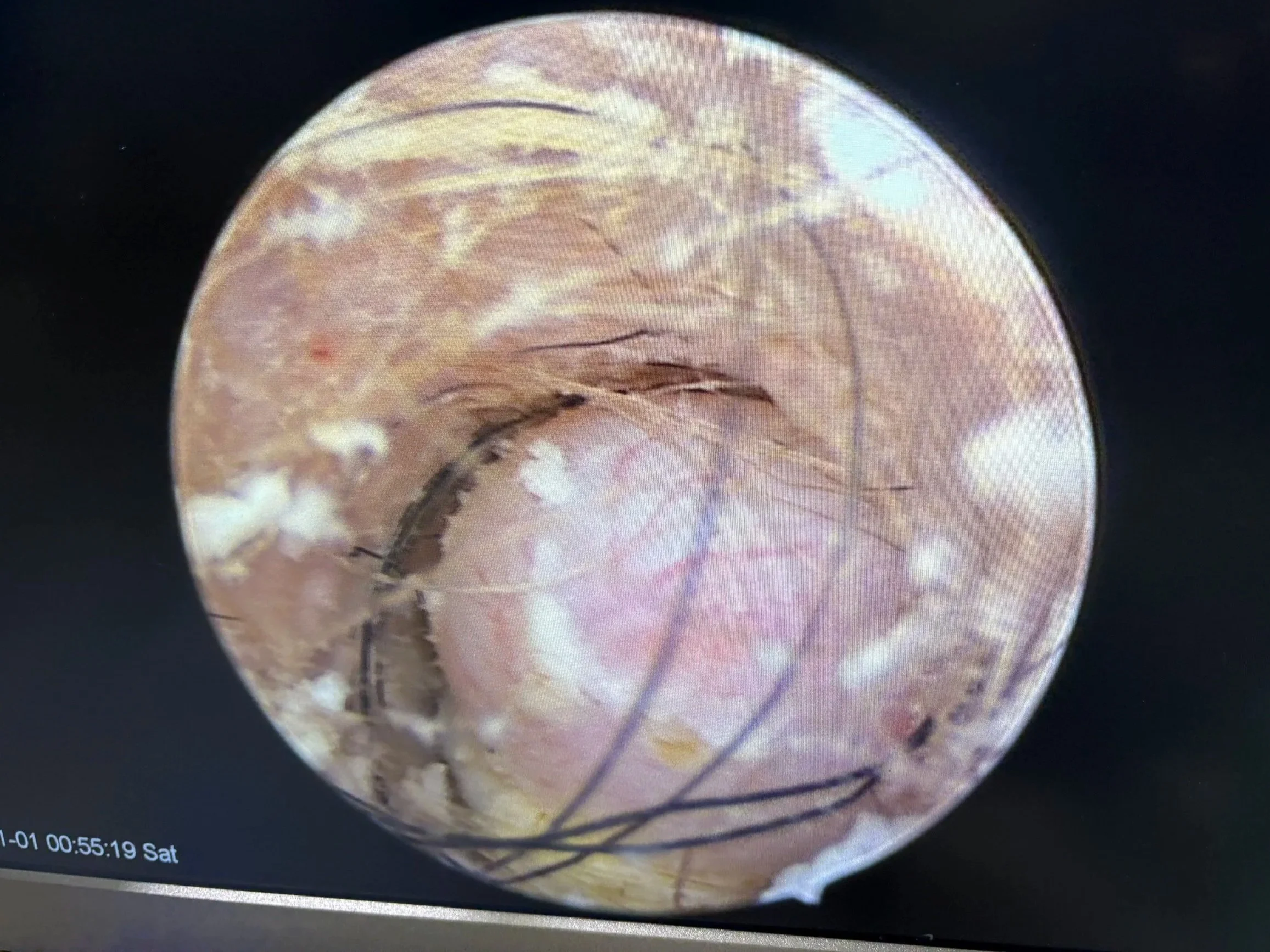
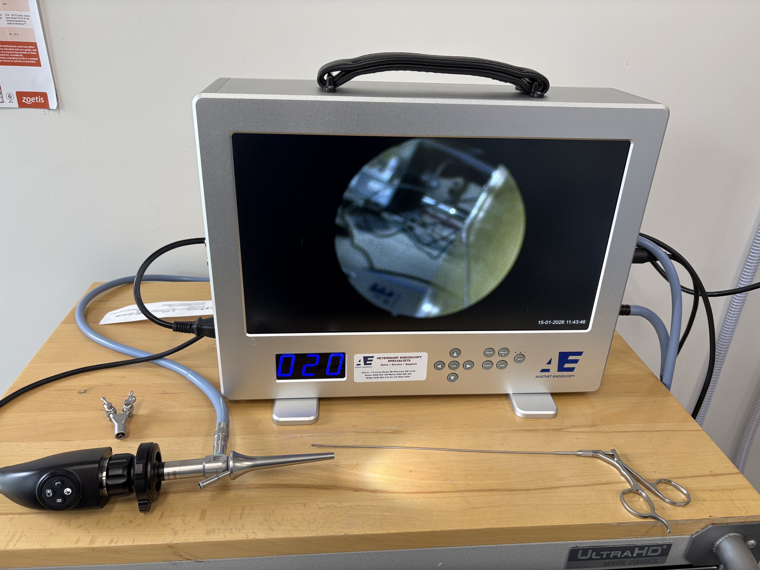
Video Otoscopy
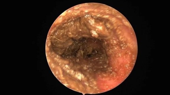
Otitis Externa
Chronic, recurrent or severe otitis externa is painful and frustrating (for your patient, their owner and you).
Often the first step towards resolution of chronic otitis externa is a thorough ear examination and deep ear clean under anaesthesia via video-otoscope. If there is significant canal stenosis, oral steroid medication is often prescribed for 1- 2 weeks to open up the ear canal prior to video-otoscopy.
Visualising and cleaning the ear canal with the video-otoscope increases the likelihood of identifying any underlying problem, or deeper complications such as ruptured tympanic membranes, otitis media, foreign bodies, ear canal masses or inflammatory polyps.
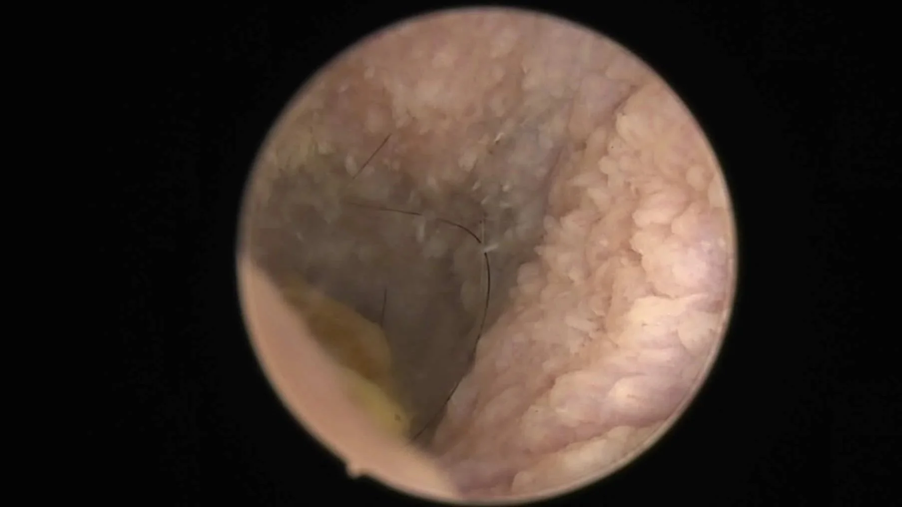
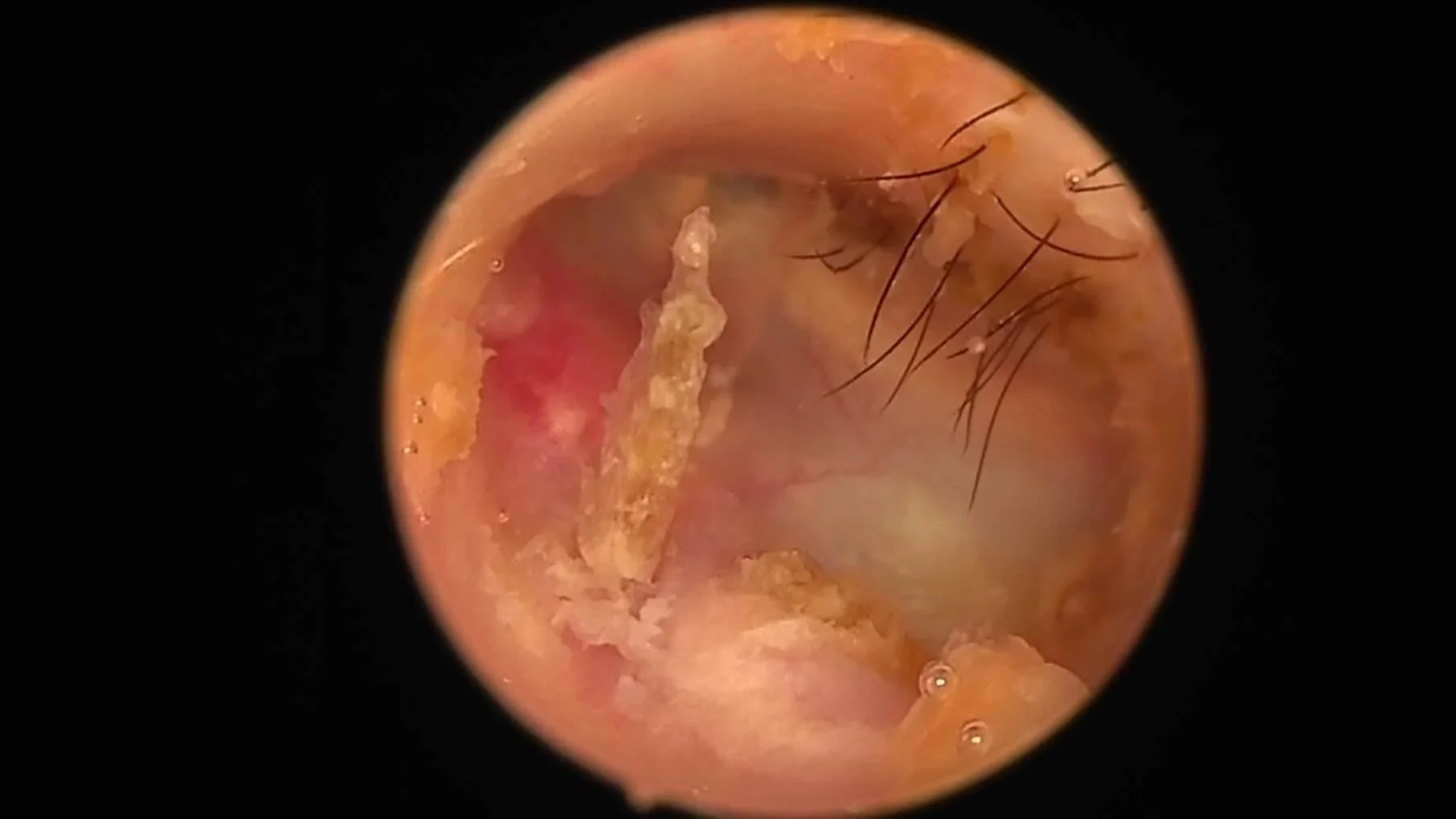
Pseudomonas Otitis
Stinky, purulent ears with rods - you can smell them in the waiting room.
These ears need a thorough deep ear clean under anaesthesia to break down and remove biofilm, remove exudate, obtain deep samples for culture and sensitivity, and evaluate the tympanic membrane.
Aggressive therapy can resolve these but follow up and investigation for the underlying cause is an important part of management and prevention of relapse.
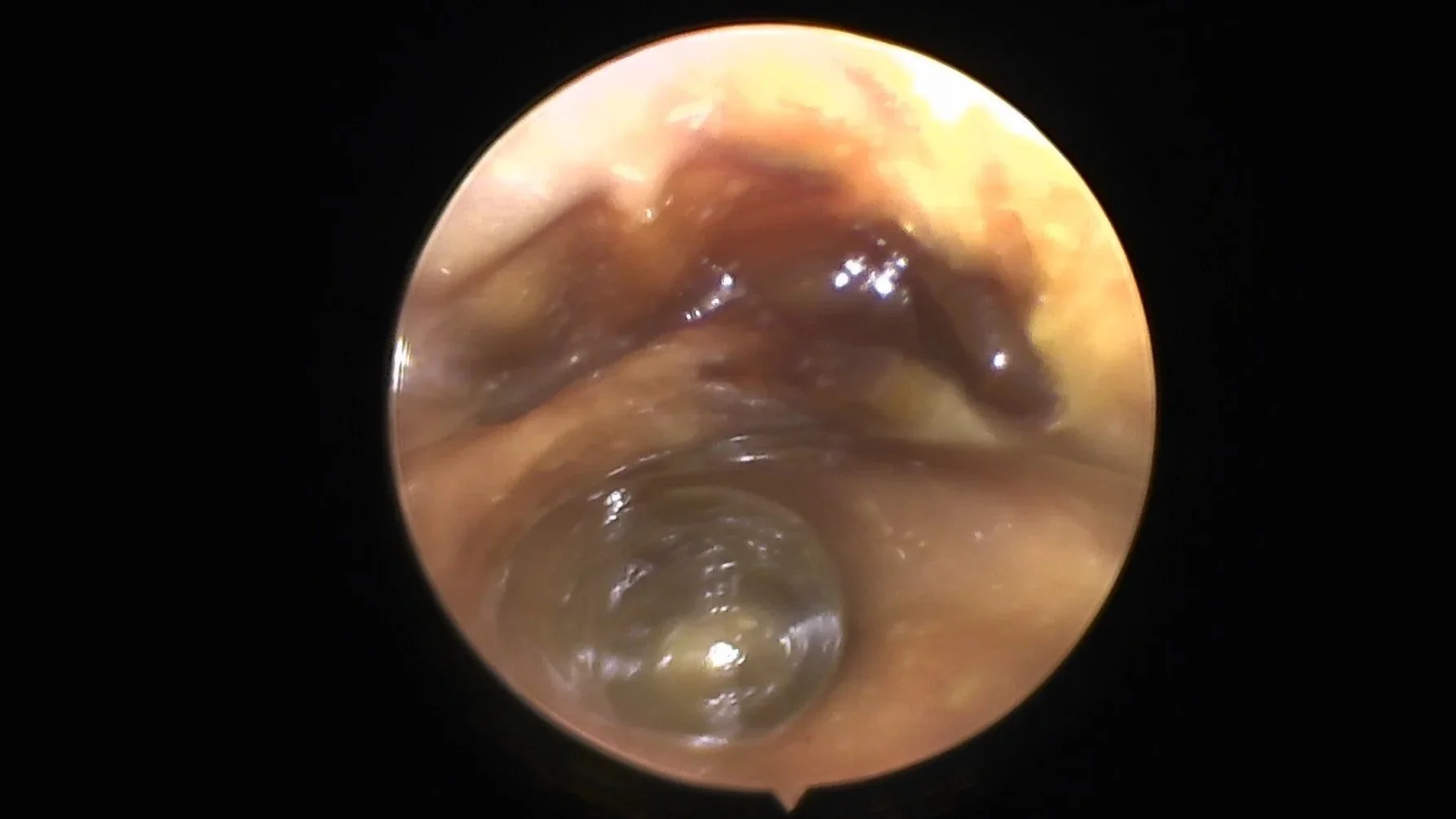
Otitis Media
We can perform a myringotomy for sample collection for culture and sensitivity, followed by a gentle but thorough lavage of the bulla. CT prior to treatment is preferred and we can arrange this if requested.
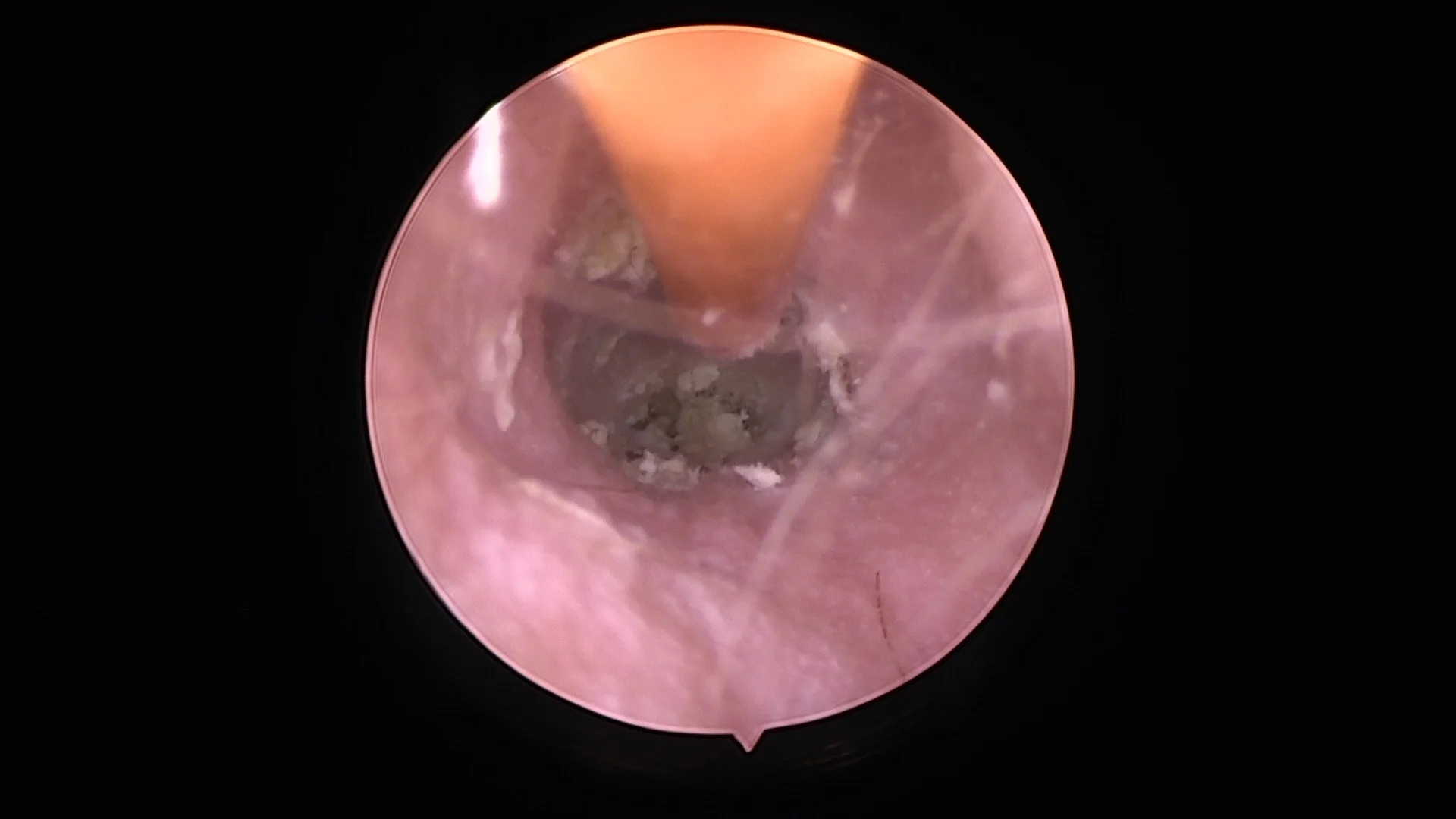
Biopsies
Ear canal masses can be identified and then safely biopsied by passing biopsy forceps through the instrument channel of the video-otoscope. Some masses or polyps can be removed via a snare.